• broken image

    Time: 29th June-3rd July 2020

    Place: Online Event

    Participation fee: 250 CHF (academia) / 500 CHF (industry)


    Update 2020-04-29: Unfortunately, ZIDAS 2020 will not take place on-site. The EPFL has decided that all campus events are suspended until August this year in order to fight against the spread of the Coronavirus. In addition, due to worldwide travel restrictions, it would be hardly possible for participants and instructors to come to Switzerland.


    In order to make the best out of the current situation, we have decided to turn this course into an online teaching event. The date will be maintained and the program will be adapted to make it compatible with online teaching!

    Best regards from the

    Organizing team


  • Anyone can apply - irrespective of affiliation, position, or location!
     

    Applications accepted from: 2020-02-01

    Application deadline: 2020-03-09 (23:59 CET)

    Application answer: 2020-04-01


    Applications are currently closed!

  • broken image

    Apply

    Anyone can apply - irrespective of affiliation, position, or location!
     
    Deadline: 15th April 2018 at 23:59 CET

    broken image

    Please support our Thunderclap campaign!

    Let us reach more people!

  • About the School

    This one-week school provides a hands-on introduction to image processing and analysis, with an emphasis on biologically relevant examples.

    Is this school for you?

    • Are you a life-science researcher with a pressing need to quantify your light-microscopy images?
    • Are you uncertain about how to: Best calculate co-localisation, do deconvolution, automate the counting of cells, track objects over time, handle massive amounts of image data, record your image-analysis workflows in a reproducible manner?
    • If you answered yes to some of the above, then this school is for you!

    Motivation

    Digital images of high quality and quantity are now the norm in biomedical sciences. Ten to twenty years ago, when many current professors trained as students or post-docs, this was not yet the case as most microscopes were, at best, equipped with low-resolution digital cameras, celluloid-film (analogue) cameras, or no camera at all.


    This rapid change is rarely reflected in the curricula of life-science university departments and they offer few, if any, courses in image processing and analysis. Understandably, courses in image-analysis (computer-vision) in the computer-science departments tend to have different aims, work on different image-data, and pre-suppose literacy in at least one programming language, rendering them all but irrelevant for the life-scientist working in the laboratory.


    To bridge this gap, between what the life-scientist needs and what courses he/she is normally offered, we have created this school for image analysis. No former experience with programming is assumed, nor will much indeed be needed; only a strong desire to learn what can be done and how to do it is required.

    What you will learn

    You will learn the fundamentals of image analysis, including basic macro programming in ImageJ/Fiji as well as other software solutions.


    MOOC

    Before the start of the course, you’re expected to validate the IPA4LS on-line course . You’ll have some videos, exercises and quizzes to gauge your IDA knowledge. We ask you to follow the “extra week” dealing with the ImageJ Macro language.


    The first day will be focused on Macro programming (from variables, to loops, functions and batch processing) while going through the concepts you discovered during the MOOC.


    In the first part of the week we will also cover the process of image-formation as it pertains to image analysis: Resolution, correct exposure, point-spread functions, detector noise, Shannon's sampling theorem, and aliasing. All with a clear focus on application in the lab.


    In the second half of the week there will be a number of focused topics, building on what was learnt during the first days, possible topics are:

    • Deep Learning for image restoration and segmentation.
    • Co-localisation, i.e., spatial correlation analysis and statistics.
    • Data Analysis using KNIME.
    • De-convolution of microscopy images.
    • Tracking of particles and cells in time-lapse recordings.
    • Stitching and registration of stacks of large image data.
    • Hands-on training with image-analysis software solutions such as ilastikCellProfiler and QuPath.

    Structure of the week

    You will be working actively with image-analysis software every day -- this is an interactive hands-on school, not a passive lecture series. Short introductions are followed by guided workflows that we step through together. You can (and should) ask questions at any time throughout.


    There will be a single invited lecture every day, alternating between scientists using image-analysis as an integral part of their biomedical research and researchers developing new image-analysis algorithms and software.


    You will have the chance to work on your own data, that you brought from home, throughout the week, with support from the trainers.


    In the second part of the week you will be working on image-analysis projects, in groups, with support from the trainers. On the last day you will present the results of your project-work.

    Factoids

    1. We will accept 25 participants --- this number is kept low to facilitate effective tutoring.
    2. The school takes place in online (changed due to COVID-19).
    3. You will need to bring your meet requirements (laptop, bandwidth, mic, etc.).
    4. Participation is only possible for the entire week.
    5. A small number of fee-waivers are available.
    6. PhD students can earn two ECTS credits from the school (we provide documents, upon approval of your local doctoral school). 
  • Program

    Expect some changes as we adapt the pace dynamically

    broken image
  • Program

    Expect some changes - the program will be updated here

  • Online Course Practical Information

    Schedules


    Based on the location of most teachers and students, know that your schedule will revolve around European time zones (9h-18h CET)

    We are aware that this might be incoenient for participants

    IT Requirements

    Participants should have access to good IT infrastructure, namely

    • a computer for working out image analysis problems
    • a good internet connection to follow the webinars and download datasets
    • a quality headset with microphone to communicate effectively.
    • A webcam

    It is recommended to have access to two screens (one for viewing live teaching material, courses and one for following along with the exercises).


    We will organize a preschool session to ensure that each one of you meets these requirements! We want to ensure a smooth experience for every participant and trainer.


    Your Own Space

    It is important that you secure a room, or secluded corner, where you can work uninterrupted for 8-10 h/day for the duration of the school.

    This is important not only for you, but also for the other participants who will often be able to see and hear you and your surroundings.

    Getting Together Online!

    Upon being selected for ZIDAS, you will gain access to a Slack channel where you will be able to discuss with the other participants!


    We will plan online social events during the week to discuss more informally and spend time together.

  • Intranet

    If you are participant, here you can find additional information

    There are no published blog posts yet.
  • Get pdf version

    Poster

    Please help us promote the event!

  • Speakers

    Scientists using or developing image analysis methods in their research

    broken image

    Professor Manley’s research focuses on automated and high-throughput super-resolution fluorescence microscopy (PALM/STORM), high-density single molecule particle tracking (sptPALM), and its application to the structure and dynamics underlying the biophysics of cells and organelles.

    broken image

    The research of my group focuses on the development of new machine learning based approaches to extract quantitative biological information from microscopy images and the design of novel computational methods to augment and improve optical microscopy. Martin is the co-creator of CARE and STARDIST.

    broken image

    The work of the group is geared fundamentaly towards medical imaging research, both in the development of new technologies and processing methods and practical clinical applications in biomedical research. Esti is one of the co-creators of DeepImageJ, a collaborative project between the BiiG at UC3M and Daniel Sage, of the BIG at EPFL.

    broken image

    The microscopy and imaging core facility supports FMI scientists and collaborators for high-end light and electron microscopy, high-content screening, X-ray crystallography, and image analysis.

    broken image

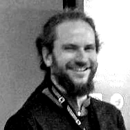
    Fabrice de Chaumont was initially hired at Pasteur to create a 3D engine to represent bio samples and image analysis results in the internal software of the unit. Then he designed a new image analysis software, Icy. His latest project involved open source cages to track complex animal behavior in real time.

  • 2018 Speakers

    (2019: Coming)

    Scientists using or developing image analysis methods in their research

    broken image

    General research topics of the lab

    • Nuclear organization
    • Intracellular transport between the nucleus and the cytoplasm
    • Nuclear pore structure and function
    • mRNA transport and degradation
    broken image

    Ivo's work focuses on developing, applying, and teaching particle methods for image-based computational biology. This includes particle methods for multi-scale simulations, bio-image processing, bio-inspired optimization, and parallel high-performance computing for particle methods. Current applications revolve around the topic of Systems Biology of Development. Ivo is the founder and head of the MOSAIC Group.

    broken image

    Our lab develops experimental and computational methods to unravel regulatory systems on the single-cell level that underlie cancer development.

    Our group’s goal is to develop methods to quantitatively analyze and model trans-cellular circuits to unravel how complex cell phenotypes in tumors are controlled. Our hope is that this will enable targeted modulation that interferes with the hallmarks of cancer and tumor development.

    broken image

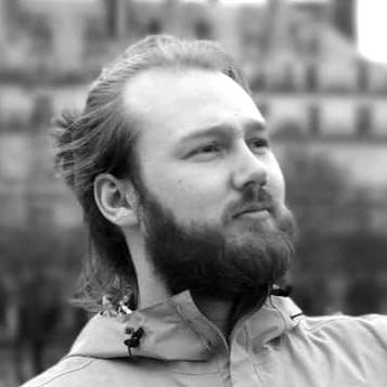
    Eleonora Secchi, from the Stocker lab, is specialized in combining microfluidics and video microscopy to study soft matter and biological samples in carefully controlled environments. She developed a new velocimetry technique called Ghost Particle Velocimetry (GPV) suitable for microscale and macroscale soft matter systems. She successfully applied this technique to study bacterial suspensions in flow.
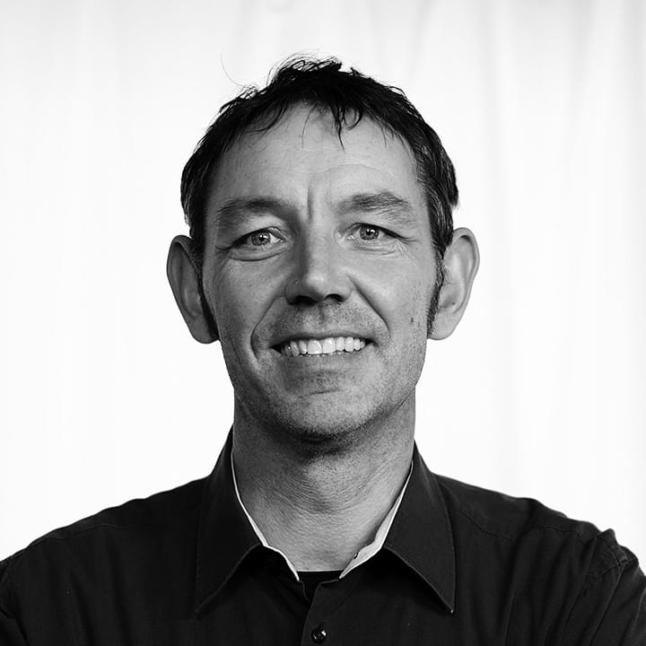
    broken image

    Dr. Kota Miura

    EMBL / University of Heidelberg, Heidelberg

    Kota Miura had been working at the Centre for Molecular and Cellular Imaging, EMBL Heidelberg as Scientist and IT engineer since 2005, and since 2014, he is working as an associate professor of the European branch office of National Institute of Basic Biology (Japan) and as a senior image analyst at EMBL. B.L.A. (Liberal Arts, ICU, Tokyo), M.Sc (Physiology, Osaka), Ph.D. (Cell and Developmental Biology, Munich). A biologist now very much specialized in image analysis.

  • Advisory Board

    SwitZerland's Image and Data Analysis School 2020

  • Connect With Us

    Something unclear? Let us know!

  • Support and Endorsements

    This school was created by ScopeM-IDA staff, co-organized with BIOP staff, powered by NEUBIAS trainers, and supported by EXCITE.

    broken image
    broken image
    broken image
    broken image
    broken image
    broken image
    broken image
    broken image
  • Sponsors

    A great thank you to our wonderful sponsors for supporting open bioimage data analysis.

    broken image
    broken image
    broken image